Structure and Function of Human Ear with Diagram Teachoo
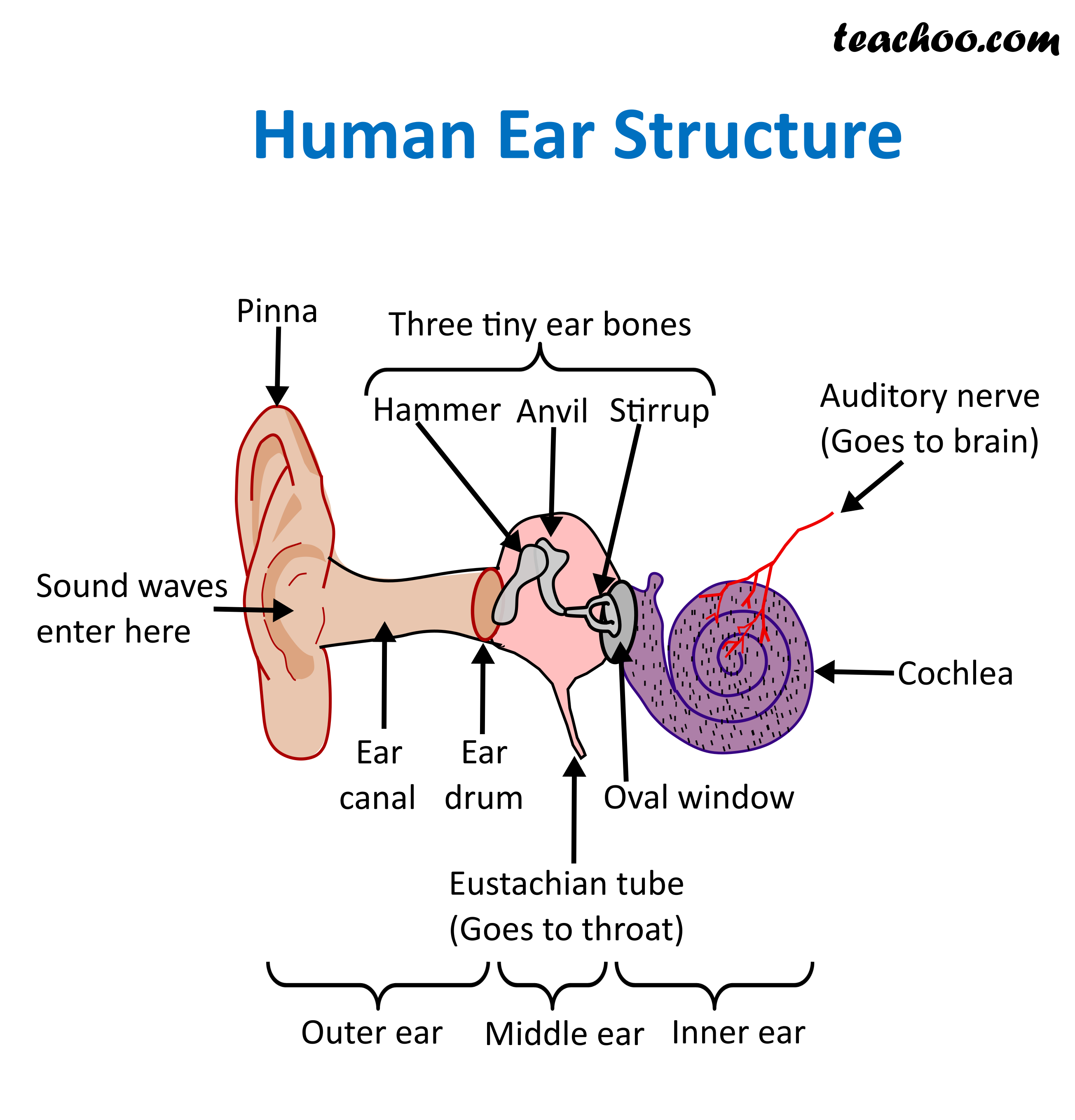
The ear is divided into three parts: Outer ear: The outer ear includes an ear canal that is is lined with hairs and glands that secrete wax. This part of the ear provides protection and.
Inner Ear Problems Causes & Treatment of inner ear Dizziness & Vertigo
Ear Anatomy | Overview & Diagram Lesson Transcript Author Anne Kamiya View bio Instructor Rebecca Gillaspy View bio Study the ear anatomy and learn about the parts of the ear. Explore.
What is a balance disorder? Hearing Link
Chapter 2 - Testing Audiogram Tympanogram Chapter 3 - Ear Anatomy Ear Anatomy - Outer Ear Ear Anatomy - Inner Ear Ear Anatomy Schematics Ear Anatomy Images Chapter 4 - Fluid in the ear Fluid in the ear Discussion Fluid in the ear Outline Middle Ear Ventilation Tubes Fluid in the ear Images Chapter 5 - Traveler's Ear Traveler's Ear Discussion
Ear Anatomy Causes of Hearing Loss Hearing Aids Audiology
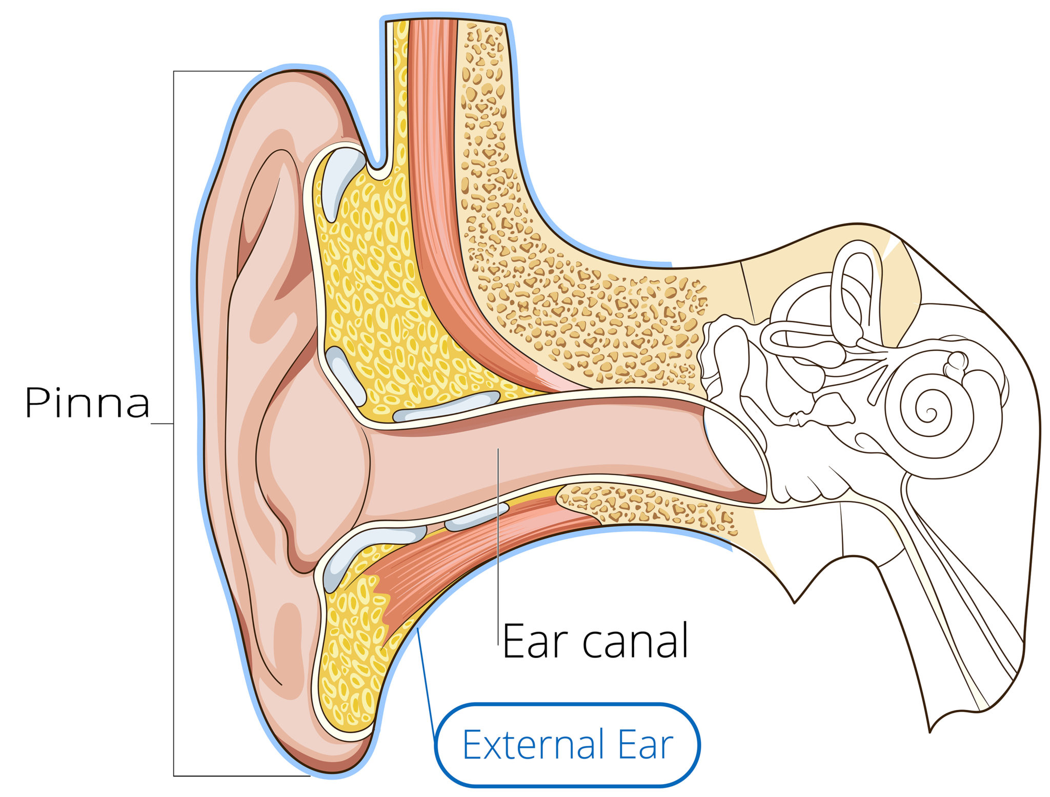
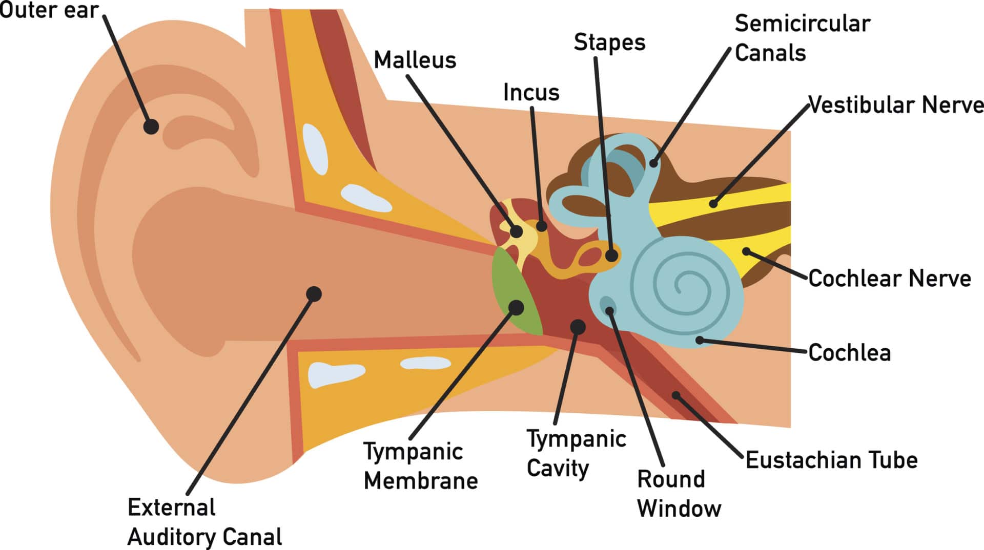
The ear canal, or auditory canal, is a tube that runs from the outer ear to the eardrum. The ear has outer, middle, and inner portions. The ear canal and outer cartilage of the ear make.
Anatomy of the Ear GomerBlog
Ear anatomy can vary. In addition to normal and relatively minor differences, there are a number of more significant and impactful variants. For instance, on the auricle, attachment—or lack thereof—of the earlobe to the face is a frequently seen genetic variation, with attached earlobes seen in anywhere from 19% to 54% of the population..
How The Ear Works Step by Step Brief Explanation
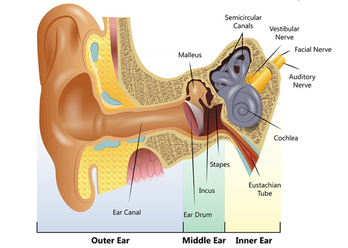
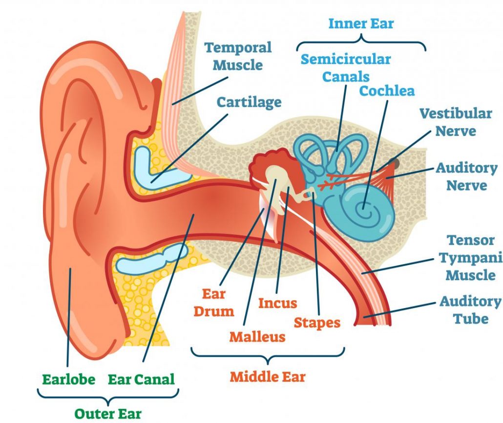
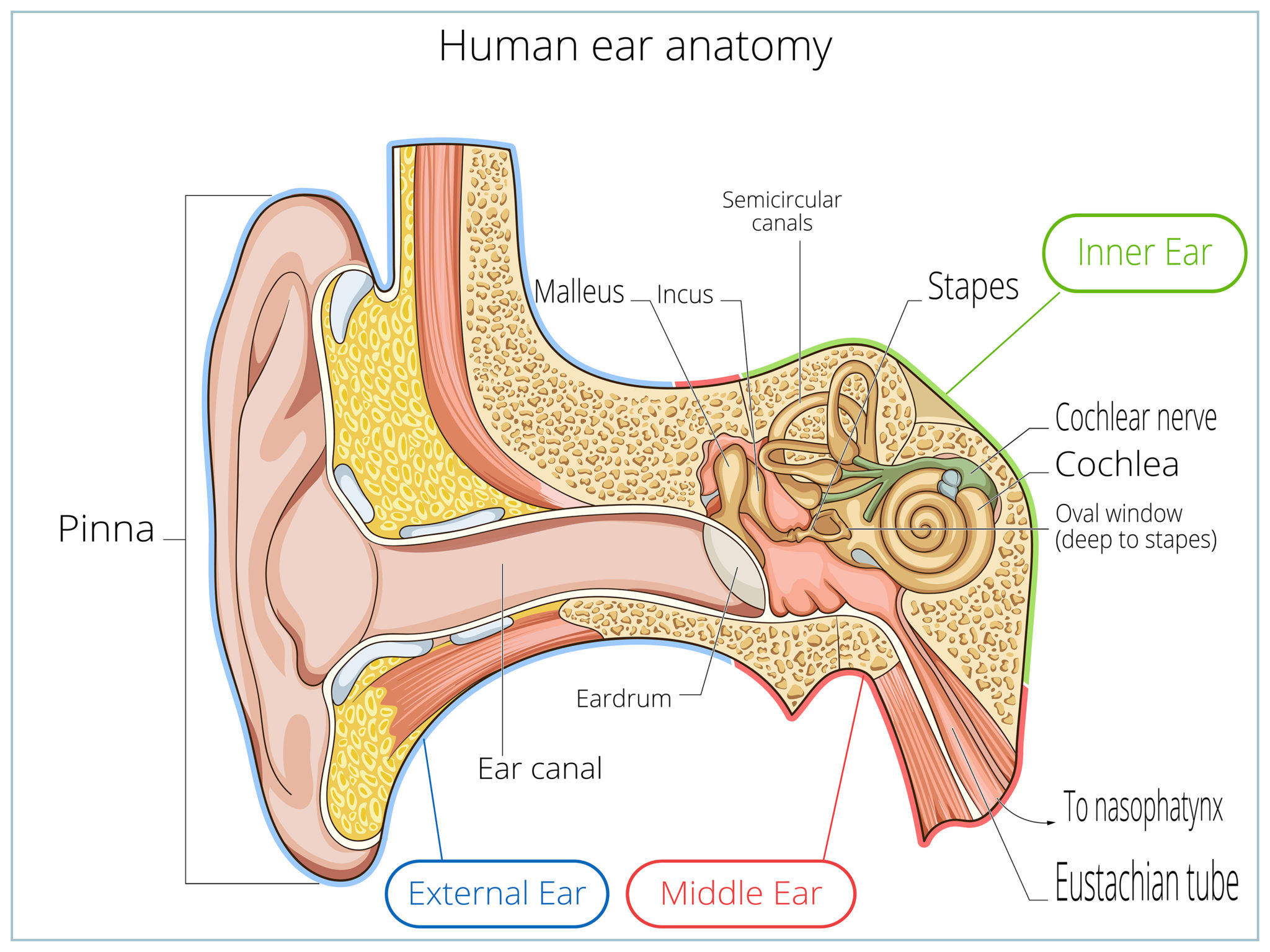
Your outer ear and middle ear are separated by your eardrum, and your inner ear houses the cochlea, vestibular nerve and semicircular canals (fluid-filled spaces involved in balance and hearing). What is the ear? Your ears are organs that detect and analyze sound. Located on each side of your head, they help with hearing and balance. Advertisement
Anatomy of the Ear
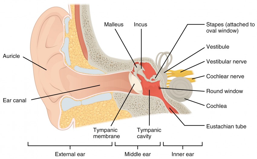
The ear is anatomically divided into three portions: External ear Middle ear Internal ear This mixture of bones, nerves, vessels, membranes, and muscles that make up the ear will be described in this article. Contents External ear Auricle External acoustic meatus Tympanic membrane Muscles of the external ear Vasculature of the external ear

How The Ear Works
human ear, organ of hearing and equilibrium that detects and analyzes sound by transduction (or the conversion of sound waves into electrochemical impulses) and maintains the sense of balance (equilibrium). Understand the science of hearing and how humans and other mammals perceive sound How humans and other mammals perceive sound.
How You Hear Northland Audiology
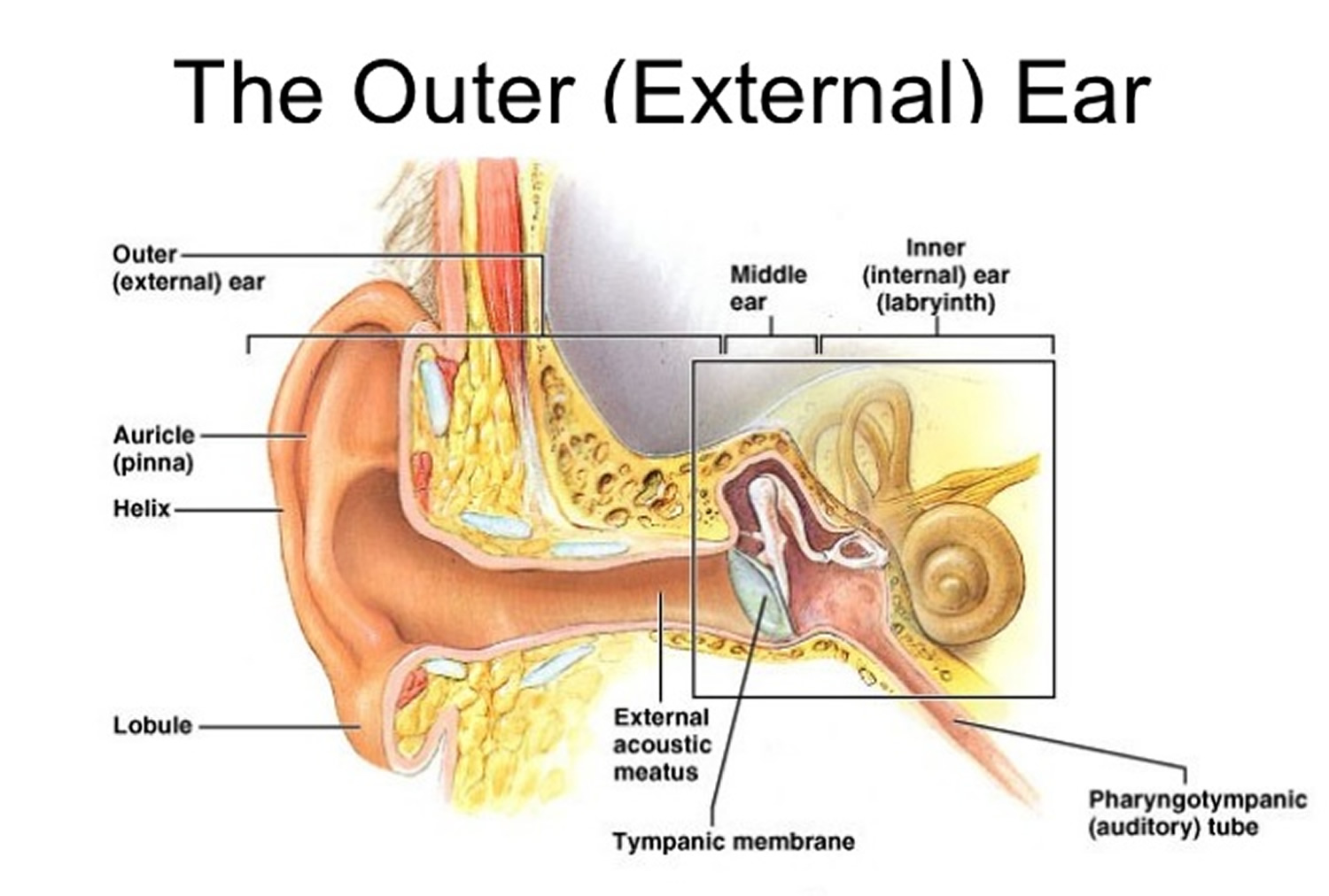
Figure 1.Anatomy of the external ear. 4 Innervation of the auricle. The auricle has several sources of sensory innervation:. The superficial surface is supplied by the great auricular nerve and lesser occipital nerve, both of which are branches of the cervical plexus (C2 & C3), and the auriculotemporal branch of the mandibular nerve, which is a branch of the trigeminal nerve (cranial nerve V)
Audition and Somatosensation Anatomy and Physiology I
So as the air vibrates even the ear drum starts vibrating. Just like the skin of a drum. And as you can, the ear drum also separates the outer ear from the middle ear. This brings us to the middle ear. The middle ear consists of the three tiniest bones of the human body. And they're together the are called the ossicles. And they have pretty.
Ear Anatomy Causes of Hearing Loss Hearing Aids Audiology
Helix: The outermost curvature of the ear, extending from where the ear joins the head at the top to where it meets the lobule. The helix begins the funneling of sound waves into the ear; Fossa, superior crus, inferior crus, and antihelix: These sections make up the middle ridges and depressions of the outer ear. The superior crus is the first ridge that emerges moving in from the helix.

EarQ Anatomy of the Ear Chart Human ear, Inner ear diagram, Ear anatomy
The ear can be divided into three parts; external, middle and inner.This article will focus on the anatomy of the external ear - its structure, neurovascular supply and clinical correlations. The external ear can be divided functionally and structurally into two parts; the auricle (or pinna), and the external acoustic meatus - which ends at the tympanic membrane.
The Ear CP Blas de Otero
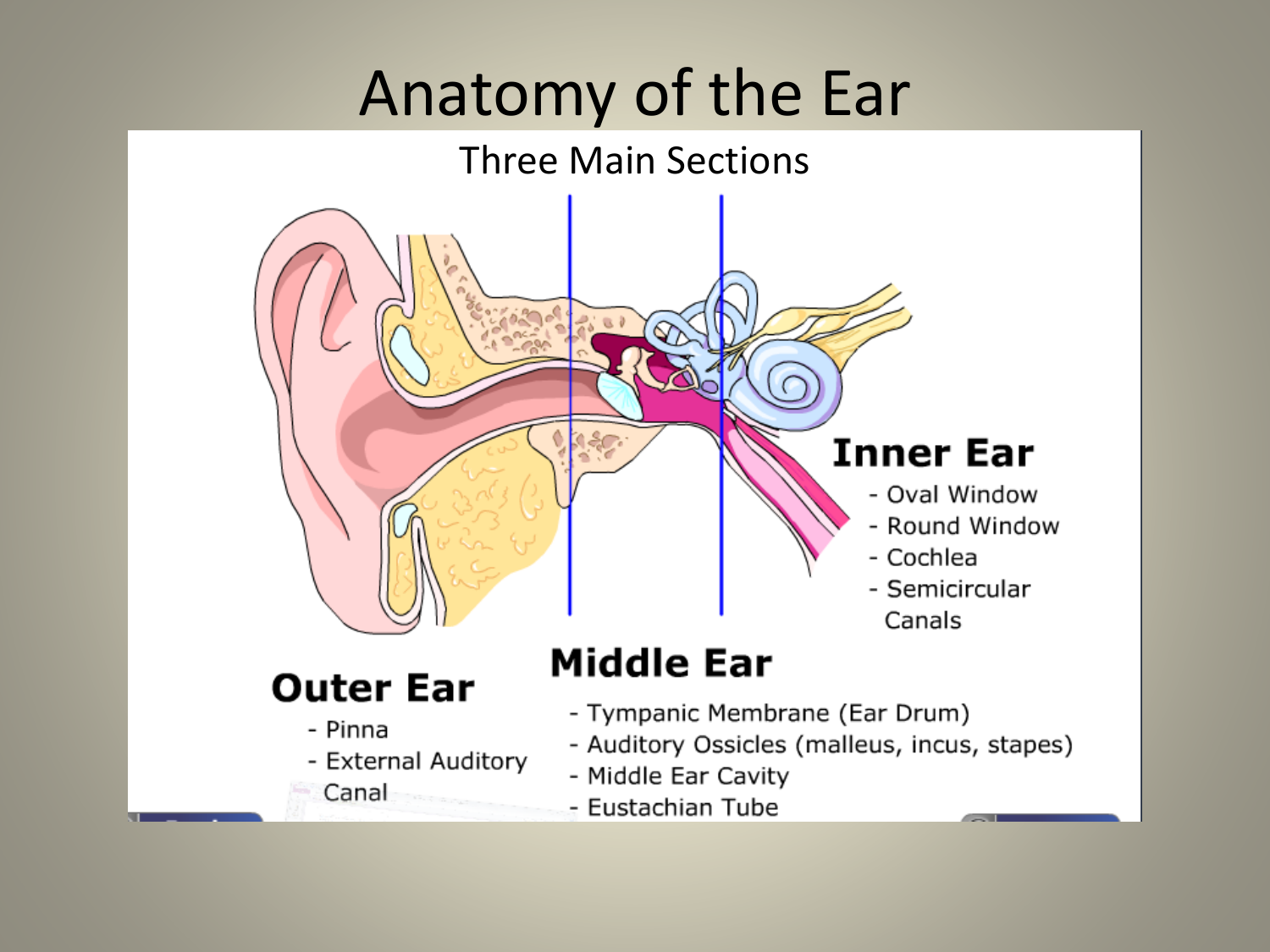
Two parts: The pinna and the ear canal Pinna: The visible part of the ear that is made of cartilage and skin Sound is transferred from the pinna to the ear canal Ear Canal: The pathway to the middle ear Earwax is made from skin glands in the ear canal Earwax protects the canal and middle ear The Middle Ear

Alila Medical Media Human ear anatomy, labeled diagram. Medical illustration
Here is a blank human ear diagram for you to label, so that you can memorize the different parts of this vitally necessary organ, for good.
Outer Ear Anatomy Outer Ear Infection & Pain Causes & Treatment
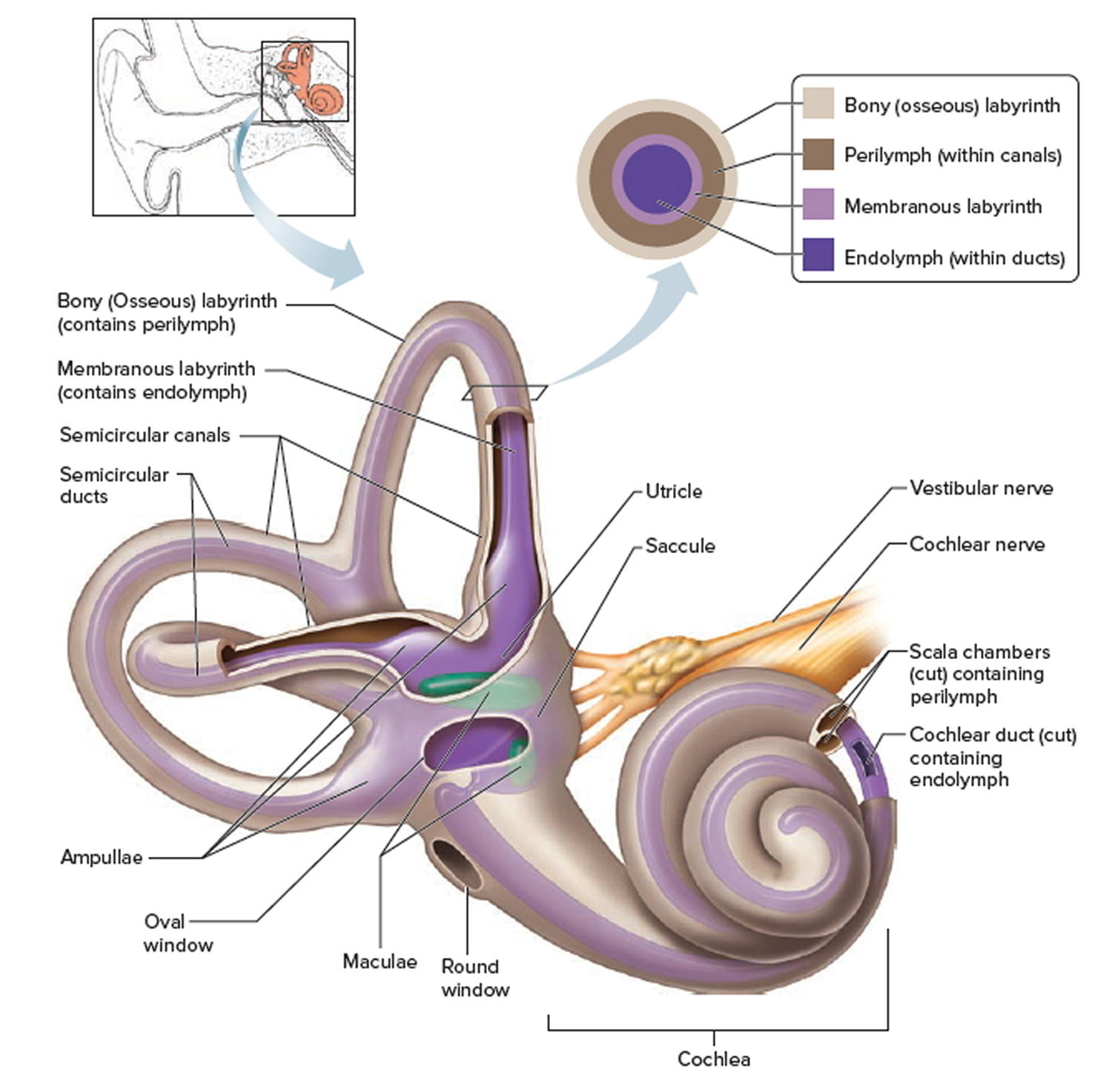
The purpose of the inner ear is to sense and process information about sound and balance, and send that information to the brain. Each part of the inner ear has a specific function. Cochlea: The cochlea is responsible for hearing. It is made up of several layers, with the Organ of Corti at the center.

HEARING ANATOMY AND PROCESS AUDIOLOGIS
Ear anatomy overview Ear diagrams (labeled and unlabeled) Accelerate your learning with interactive quizzes Sources + Show all Ear anatomy overview Although it's not obvious to look at, the ear is anatomically divided into three portions: External (outer) ear Middle ear Inner ear